Meet us at the ICC2025 in Florence!

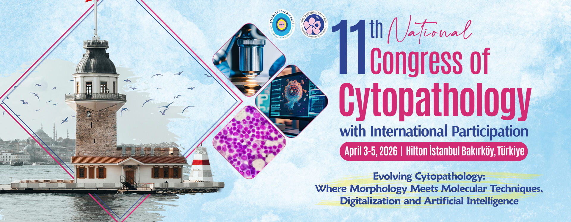
Dear Friends and Colleagues,

We are quickly approaching the International Cytology Congress in Florence—an outstanding opportunity to immerse ourselves in all that cytology has to offer today. With a rich scientific program designed to broaden our knowledge, it will also be a perfect occasion to exchange ideas, make new friends, and reconnect with old friends.

In this global gathering, there will be many chances to engage with the EFCS family as well. On Sunday, 11th May at 11:00, Cavaniglia Pavillion Room A, join us for the EFCS Companion Meeting, where you’ll hear about exciting educational and scientific initiatives within the EFCS, chaired by Danijela Vrdoljak Mozetic and Ambrogio Fassina.

The session will start with Ivana Kholova, who will set the stage by reflecting on the past and present to help us envision the future. Esther Diana Rossi will then guide us through the world of interventional cytology, followed by Claire Burgain, who will share insights into the current state of cervical screening. Irena Srebotnik Kirbis will present her comprehensive work on cell block techniques, and Pawel Gajdzis will introduce the ASCUS Young Project—an EFCS initiative with involvement of early-career cytologists. Finally, I’ll have the pleasure of representing the Young EFCS, sharing updates on our training and educational projects.

A special highlight will be the Cytology Social Media Agora—a true novelty not to be missed on the 13th May starting at 11:15, Cavaniglia Pavillion Room B. As our professional lives are increasingly shaped by social media—for networking, education, and consultation—Cioly Rivero Colmenarez will represent us from Europe in this exciting session.

But that’s not all! The Young EFCS will also be present at the Young EFCS Booth at the Congress. Come by to meet us, learn more about our work and projects, and—if you feel inspired—join our rapidly growing community.

Looking forward to seeing you all soon!

Warm regards,
Istvan Kovacs
Young EFCS

Read More »

Case of the Month March – by Ena Holjevic

Clinical Presentation:

An 82-year-old male patient arrived at the emergency department with shortness of breath, a dry cough that improved with rest, and swelling of his left leg. A CT pulmonary angiography was performed to rule out pulmonary embolism, revealing no signs of thrombosis; however, bilateral pleural effusions were detected. Additionally, a CT scan of the abdomen and pelvis was performed, but no tumor mass was found. The clinicians performed a therapeutic pleural puncture and sent the pleural fluid for cytological analysis.

A cytological smear was prepared from the pleural fluid sediment. Microscopic examination revealed numerous large atypical lymphoid cells with irregular nuclear contours, prominent nucleoli, and abundant basophilic cytoplasm. Some cells displayed plasmacytoid features with occasional vacuolization.

Highly cellular pleural fluid sediments showed large atypical lymphoid cells with prominent nucleoli and abundant cytoplasm.

High magnification view highlighting plasmacytoid morphology and mitotic activity.

Prominent nucleoli, abundant basophilic cytoplasm.

Cytoplasmic vacuoles, multinucleation.

A cell block was prepared from the pleural fluid sediment for further analysis.

Immunohistochemical (IHC) staining was performed on the cell block sections to reveal the immunophenotype of the atypical lymphoid cells. The tumor cells were negative for CD20 and CD3, while showing positive staining for CD30, CD138, HHV-8 and MUM 1.

CD3 (200x magnification): Demonstrating T-cell marker expression.

CD20 (200x magnification): Demonstrating B-cell marker expression.

CD30 (400x magnification): Demonstrating variable CD30 expression in lymphoma cells.

CD138 (400x magnification): Demonstrating plasma cell differentiation marker.

HHV-8 (400x magnification): Demonstrating HHV-8 positivity in tumor cells.

MUM1 (400x magnification): Demonstrating MUM1 expression in tumor cells.

Flow cytometry: Analysis of the cells within the lymphocyte gate shows 72% T lymphocytes and 3% B lymphocytes. The B cells are mature monoclonal B cells (CD19+CD20+CD22+) that express lambda light chains on their surface (kappa/lambda ratio: 1.2; normal range = 0.5-

4.0). The T cells are of mature phenotype and have a normal ratio of helper and cytotoxic T lymphocytes (CD4/CD8 = 1.0; normal ratio = 0.9-3.4). The majority of cells in the sample (60%) are mononuclear cells of the phenotype CD45+CD19-CD3-CD38+CD138+, which most closely corresponds to plasma cells.

A bone biopsy and puncture were also performed, revealing no evidence of infiltration from the underlying disease.

Antibody testing was conducted for Epstein-Barr virus, cytomegalovirus, human immunodeficiency virus, and hepatitis B and C. The test results were positive for EBV IgG and CMV IgG.

Discussion:

This case highlights the importance of considering Primary Effusion Lymphoma in the differential diagnosis of unexplained pleural effusions, particularly in elderly or immunocompromised patients. Primary Effusion Lymphoma is a rare and aggressive B-cell non-Hodgkin lymphoma associated with Human Herpesvirus 8 (HHV-8) infection. It is typically seen in immunocompromised patients, particularly in those with HIV/AIDS, but can also occur in elderly individuals with age-related immunosenescence. The differential diagnosis includes malignant mesothelioma, metastatic carcinoma, and other lymphomas (such as large B-cell lymphoma with plasmablastic features). The absence of CD20 expression and positivity for HHV-8 are key distinguishing features.

Key Learning Points:

  1. PEL presents as a malignant effusion without a detectable tumor mass.
  2. HHV-8 positivity is a defining feature and aids in diagnosis.
  3. It primarily affects immunocompromised individuals but can also occur in elderly patients.
  4. The prognosis remains poor despite chemotherapy, highlighting the need for novel therapeutic approaches.

Author of the case:

Ena Holjević, MD; Special thanks to assistant professor Christophe Štemberger,MD, PhD and assistant professor Irena Seili-Bekafigo, MD, PhD for their support in the developing of this case.

References:

Cesarman, E., Mesri, E. A., & Gershengorn, M. C. (2023). Primary Effusion Lymphoma. Diagnostics, 13(3), 370.

Coyle, T., Xie, Y., & Shergill, A. (2021). Primary Effusion Lymphoma: Current Perspectives. Frontiers in Oncology, 11, 33669719.

Read More »

Diagnostic Procedures in Patients With Neck Masses

Jerzy Klijanienko,

Beatrix Cochand-Priollet,

Olivier Choussy,

Wojciech Golusinski

(Editors),

Springer Nature, 2024.

ISBN: 9783031676741

The ENT region is of great anatomical complexity. The proximity of tissues of different origin, a specific clinical terrain and the presence of organs exposed to viruses and other aggressive agents mean that this region presents a great variety of lesions of different nature: inflammatory, benign tumors and malignant tumors can present overlapping clinical, radiological and pathological characteristics.

In addition, this richness (diversity) of presentation is the source of “hyperspecialization” in the involved medical specialties. There are specialists for the thyroid gland, salivary glands, connective tumors or, finally, the upper aero-digestive tract. As much as some chapters of this pathology can be quite common, others can be extremely rare and therefore more difficult to diagnose.

There is a simple explanation for this: the diagnosis of rarer, more complex and more difficult lesions is more easily managed by centers specialized in this field. In an attempt to cover this complicated field of pathology, we have invited leading specialists engaged in diagnosis to cooperate.

First, the former President and the former General Secretary of the European Federation of Cytology Societies, the former President of the European Head and Neck Society and the current head of oncological surgery at the Institut Curie in Paris represent the four Editors. The introduction was written by Prof Andrew S. Field, from the University of New South Wales, former President of the International Academy of Cytology as well as Prof Fernando Schmitt, from the Medical Faculty, University of Porto, current president of the International Academy of Cytology, both widely implicated in the IAC WHO IARC Joint Standing Editorial Board for the publication of the so-called “Blue Books” (i.e international systems for reporting cytologies).

The chapters were entrusted to specialists in the different fields coming from numerous Centers and prestigious universities. Chapters in clinical, radiology, nuclear medicine, virology, immunohisto/cytochemistry, molecular biology, dermatology, and ophthalmology offer an original scientific approach of these Head and Neck lesions. In seeking the objective of clinical objectivity and usefulness, we treated classical pathology and cytopathology techniques equally. Scientific arguments in favor of one technique or the other were shown and discussed. We are aware that different oncological centers have their own diagnostic methods and we tried to unite them, discussing and showing the advantages of other experiences.

In hopes that all of this will serve our patients and that they will benefit from modernity and efficiency in medicine in 2025.

Prof J Klijanienko

Ass Prof B Cochand-Priollet

Dr Olivier Choussy

Prof W Golusinski

Read More »