European Congress of Cytology 2026
After 24 years Belgium again has the honour to welcome a distinguished company from Europe and beyond for the 46th European Congress of Cytology. Back then for a young resident it was the first big international congress and the experience was incredible. So it will be again, when we meet old and new friends, learn from each other and grow together.
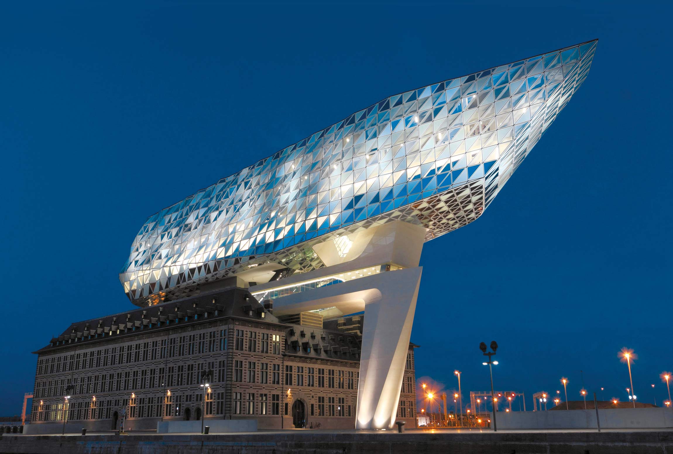
Antwerp has a long history in the diamond trade and knows the meaning of brilliance. It is only natural that it will host an outstanding cytology congress. The discipline is stepping into the digital age and will shine brighter with use of new tools that come with it. Some of the brightest and most brilliant minds in our community will show us what this future can offer. AI assisted diagnoses are already possible. Digital cytology will help us to work more efficiently. Ancillary techniques, improved classification systems and patient tailored treatments will yield better patient management. This is all possible with minimally invasive cytology.
Antwerp is an old and distinguished city and so is our profession. But we all know it is the new insights and the new, young colleagues, that give it its long life. We all joined a global, brilliant community, which shares the passion for cytopathology. We are happy to be able to share this experience with friends, old and new alike.